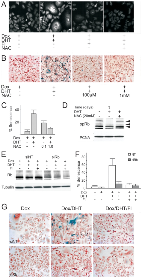
Figure 4. AR-induced senescence requires ROS and Rb.
This figure depicts the role of ROS and RB in AR-dependent senescence. (A). PC3-AR cells were treated with Dox, DHT and Fl, as indicated and ROS assessed by DHE fluorescence. Note increased ROS upon DHT treatment and lower DHE fluorescence in the presence of Fl (20 µM). ROS quencher, NAC (10 mM) was used as a negative control. (B, C) The effect of NAC on AR-induced senescence: PC3-AR cells were cultured in Dox, AR activated with DHT ± NAC, and senescent cells visualized with SA-βGal assay. Representative images (B) and quantitative assessment (C) are shown. (D) PC3-AR cells were cultured in Dox, treated 24 hours with DHT and/or NAC (20 mM) where indicated. Cell extracts were analyzed by western blot for phospho-Rb. PCNA was used to assess loading. Note increased Rb phosphorylation in the presence of NAC. (E) PC3-AR cells were transfected with Rb siRNA and Rb levels measured after 24 hours by western blot. Note ∼2-fold reduction after transfection with Rb siRNA. (F, G) Senescence was measured by SA-βGal assay on day 6 after Rb knock-down (values calculated as in Fig. 1, S.D. values are shown; P<0.0002). Note the reduced senescence of the DHT-treated cells after Rb knock-down. Representative images are shown below (G).

