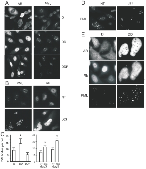
Figure 6. AR alters PML aggregation via p63 pathway.
PML tumor suppressor was linked to AR dependent senescence. (A, C) PC3-AR cells were cultured in Dox (D) and treated with DHT (DD) or DHT and Fl (DDF) for 48 hours and stained for AR, to assess activation state, and for PML (A). The number of PML bodies per 20× field was measured using MetaMorph software and recalculated per single cell (C, left). Note a significant increase in the number of PML bodies/cell in the presence of DHT (P<0.02), but not in the presence of Fl. (B, C) PC3-AR cells were transfected with NT or p63 siRNA, cultured for 3 and 5 days and stained for PML and Rb. The number of PML bodies per cell was calculated as above (C, right). Note a robust decrease in PML staining upon p63 knock-down (day 3, P<0.05; day 5, P<0.0001). (D) PC3-AR cells were transfected with non-target (NT) or p21 siRNA, treated for 3 days with DHT and stained for PML. Note decreased staining upon p21 knock-down. (E) PC3-AR treated 3 days with Dox ± DHT and stained for AR, Rb and PML. Note AR activation and nuclear translocation, concomitantly increased intensity of Rb staining with appearance of partial aggregation, and co-localization of Rb aggregates with PML nuclear bodies.

