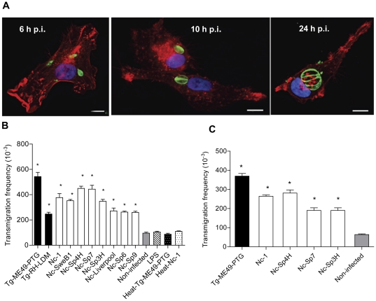
Figure 1. In vitro migratory phenotypes of DC infected with N. caninum and T. gondii.
A. Human monocyte-derived DC are permissive to N. caninum. Immunofluorescence staining of DC (phalloidin-Alexa Fluor 596) infected with the isolate Nc-1Luc (MOI 3). Parasites were labelled with hyperimmune rabbit antiserum and Alexa Fluor 488 as secondary antibody. Overlay with DAPI (blue). Representative images of 6, 10 and 24 h p.i. are shown. Scale bar: 10 µm. B. Bar diagram shows the transmigration frequencies of human monocytic-derived DC incubated with live tachyzoites (MOI 2) from T. gondii (Tg-RH-LDM and Tg-ME49-PTG strains) and N. caninum (Nc-1, Nc-Liverpool, Nc-SweB1, Nc-Spain 3H, Nc-Spain 4H, Nc-Spain 6, Nc-Spain 7 and Nc-Spain 9 isolates), CM (non-infected cells), LPS (100 ng/ml) and heat-inactivated parasites (heat-Tg-ME49-PTG or heat-Nc-1) as indicated under Materials and Methods. The black bars indicate T. gondii and the white bars indicate N. caninum. Cell migration (migrated/added) was assessed by optical counting of transmigrated cells across a transwell filter. Means (±SEM) from the replicates from at least three independent experiments are shown. Asterisks indicate treatments which conferred significant differences in transmigration compared with non-infected cells (P<0.001; Dunnett's test). C. Transmigration of BALB/c bone marrow-derived DC infected with T. gondii (Tg-ME49-PTG, MOI 2) or N. caninum (Nc-1, Nc-Spain 4H, Nc-Spain 7 and Nc-Spain 3H, MOI 2) as indicated under Materials and Methods. Means (±SEM) from the replicates from at least three independent experiments are shown. Asterisks indicate significant differences compared to non-infected cells (P<0.05; Dunnett's test).

