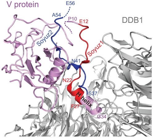
Figure 7. Structure of the V protein from parainfluenza virus 5 bound to DDB1.
The PDB accession number of the structure is 2HYE. Aa 1–9 and aa 55–80 of V, encompassing the last 2aa of soyuz2, are not visible in the crystal structure, presumably because they are disordered (see text). Soyuz1 is coloured red and soyuz2 blue. The H helix of V, bound to DDB1, is indicated; it partially overlaps with soyuz1.

