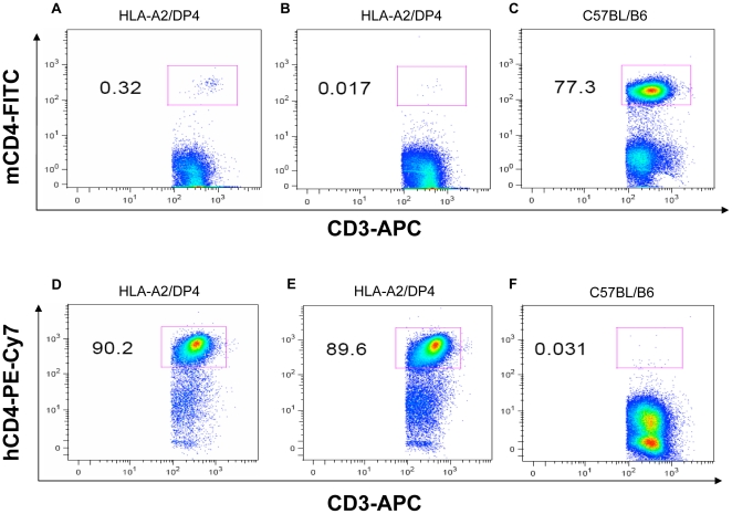
Figure 3. Flow cytometric analysis of peripheral mCD4+ and hCD4+ T lymphocytes.
Splenocytes from HLA-A2/DP4 (Figure 3A, 3B, 3D and 3E) and wild-type C57BL/B6 (Figure 3C and 3F) mice were isolated and CD3+ T cells were gated by staining with APC-conjugated anti-CD3 mAb. Meanwhile, FITC-conjugated anti-mCD4 and PECy7-conjugated anti-hCD4 antibodies were simultaneously used to observe the mCD4 and hCD4 expression in HLA-A2/DP4 and WT C57BL/B6 mice.

