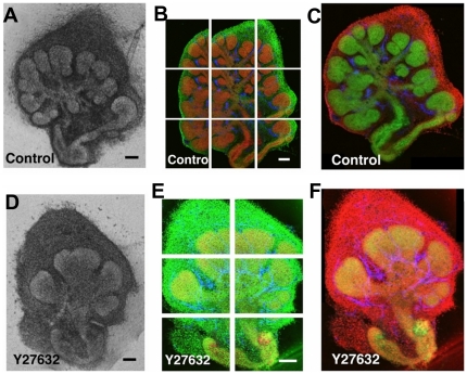
Figure 1. Acquisition and image processing of confocal images.
Organotypic culture of E13 SMGs (a) control or (b) treated with ROCK inhibitor (140 µM Y27632), showing reduced branching with ROCK inhibitor treatment. Explants were immunostained with anti-E-cadherin antibody as an epithelial marker (red) and SYBR green as a total nuclei marker (green). Multiple overlapping confocal images through the mid-section of (c) control- and (d) ROCK inhibitor-treated explants were captured to cover the whole explant. Images were stitched using the inverse Fourier transform of the phase correlation matrix and blended to provide composite images of (e) control (f) and ROCK inhibitor treated explants. Scale bars: 200 µm (a, b), 100 µm (c), (d), and (e), and (f). In our study, the sublingual tissues were discarded and only the submandibilar gland was used, (Figure S2).

