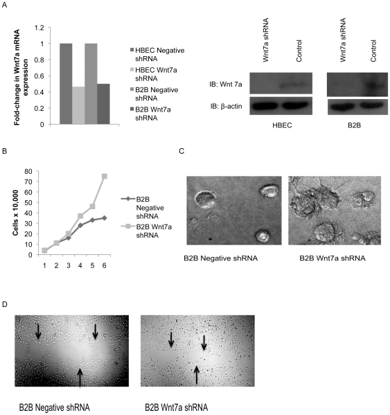
Figure 1. Effects of Wnt7a loss in lung epithelial cells.
A) Expression of Wnt7a was measured by QPCR in HBEC and B2B cells after stable transfection with Wnt7a shRNA or a negative shRNA. Data is presented as fold-change in Wnt7a expression normalized to GAPDH. Error bars are the SE of triplicate experiments. Wnt7a knock down was also confirmed by western blot with a β-actin loading control. B) Cell proliferation was measured in B2B cells expressing Wnt7a shRNA or a negative shRNA. Cells were analyzed in a 6-day growth assay. C) B2B cells with Wnt7a shRNA were analyzed in a 3D culture assay using Matrigel and observed after 5 days of growth for changes in morphology compared to a negative shRNA control. B2B cells with Wnt7a shRNA were also analyzed in a scratch migration assay and observed at 24 hours for movement into the introduced scratch compared to a negative shRNA control. Arrows indicate edges of the scratch. Pictures represent triplicate experiments.

