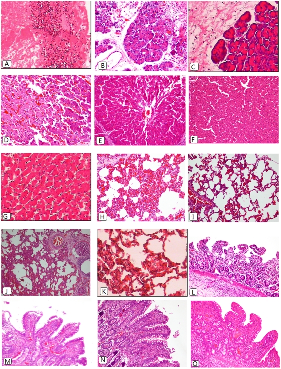
Figure 4. Histomorphological images of pancreas (A–C), liver (D–G),lung(H–K) and ileum (L–O) of SAP+IAH animals (A,D,H,L),SAP animals(E,I,M), IAP 30 mmHg animals(B,F,J,N) and sham-operated animals(C,G,K,O).
Remnant necrotic tissue with a great number of inflammatory cells infiltration(A×200).Mild-to-moderate-grade edema and degenerative changes with very mild necrosis(B×200). Mild edema and inflammation without evidence of necrosis(C×200). Livers of the study groups (D×400) showed more severe congestion and necrosis when compared with the SAP group(E×200),while there were mild-to-moderate-grade congestion, vacuolization and leukocyte infiltration in IAP 30 mmHg group(F×200). Only minor changes could be observed in the sham controls (G, 400×). The lung of the SAP+IAH animals displayed interstitial infiltration, focal hemorrhage and atelectasis(H×200) while the specimen of SAP animal presented lower grade injury(I×200). The lung specimen of IAH alone animals displayed atelectasis as well, but only moderate hemorrhage and interstitial infiltration(J×200). Slight edema and infiltration were seen in sham animals(K×400). The small bowel specimen of the study group (L×200) displayed denuded villi. The specimen of SAP animals presented extended subepithelial space and lymph follicle hyperplasia(M×200), while in the IAH group, there was similar histological changes to the SAP group and also lymph follicle hyperplasia(N×200). In the sham control, only minor changes could be seen(O×200).

