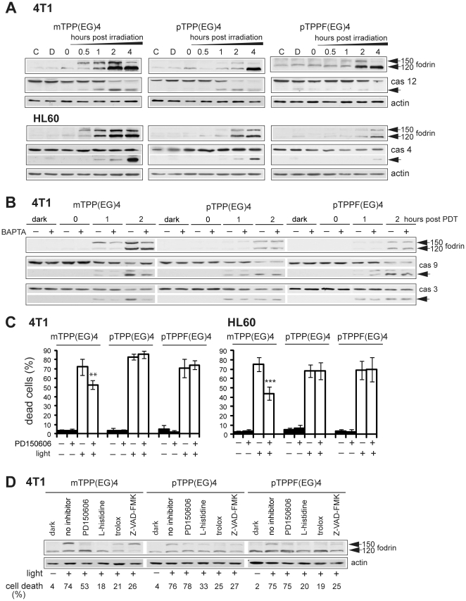
Figure 6. Effect of EG-porphyrin derivatives on Ca2+ signaling pathway.
(A) Western blot analysis of fodrin, caspase-12 (4T1 cells) and caspase-4 (HL60 cells). Cells treated with EG-porphyrin derivatives were harvested at various times after irradiation and subjected to Western blot analysis with antibodies recognizing fodrin and caspase-12. Reprobing with β-actin antibody was used to confirm equal loading. The arrowheads point to activated forms. (B) Pretreatment of 4T1 cells with BAPTA-AM (10 µM) resulted in the inhibition of fodrin, caspase-9 and caspase-3 activation caused by mTPP(EG)4-PDT. 4T1 cells loaded with porphyrin derivatives were pre-incubated for 2 h with BAPTA-AM and then exposed to light (2.5 Jcm−2). At various times after irradiation the cells were lysed and analyzed with antibody recognizing fodrin, full-length and cleaved p39 form of caspase-9 and full-length and cleaved p17 form of caspase-3 on Western blots. (C) Effect of calpain inhibitor PD150606 on viability of 4T1 and HL60 cells. Cells were incubated with PD150606 (20 µM) for 1 h and then irradiated. Cell viability was estimated after 24 h by the trypan blue exclusion method. The percentage of dead cells was expressed as the mean ± SD (n = 4–5). **P<0.01, ***P<0.001 represents statistical differences between PDT-treated cells vs. PDT-treated cells in the presence of PD150606. (D) The effect of calpain inhibitor, ROS scavengers (L-histidine and trolox), and caspase inhibitor (Z-VAD-FMK) on the activation of fodrin. Cells were treated as described in Materials and Methods, harvested 1 h post PDT treatment and analyzed by Western blot. Equal protein loading is demonstrated by actin reprobing. The viability of cells subjected to simultaneous treatment done in parallel is presented under the Western panel.

