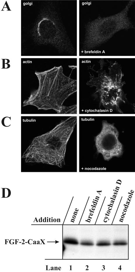
Figure 4.
In vivo prenylation of FGF-2-CaaX is not blocked by treatment with brefeldin A, nocodazole and cytochalasin D. (A) NIH/3T3 cells were treated without or with 2 μg/ml brefeldin A for 30 min. The Golgi apparatus was visualized with anti-β-COP and Cy3-conjugated anti-rabbit antibody using confocal microscopy. (B) NIH/3T3 cells were treated without or with 10 μg/ml cytochalasin D for 30 min. Actin was visualized with rhodamine-conjugated phalloidin. (C) NIH/3T3 cells were treated without or with 33 μM nocodazole for 30 min. Microtubuli were visualized with antitubulin and rhodamine-conjugated anti-mouse antibody. (D) NIH/3T3 cells were pretreated as in Figure 2A and then treated for 6 h with heparin and FGF-2-CaaX in the absence (lane 1) or presence of 2 μg/ml brefeldin A (lane 2), 10 μg/ml cytochalasin D (lane 3), or 33 μM nocodazole (lane 4). After lysis, cellular material was analyzed as in Figure 2A.

