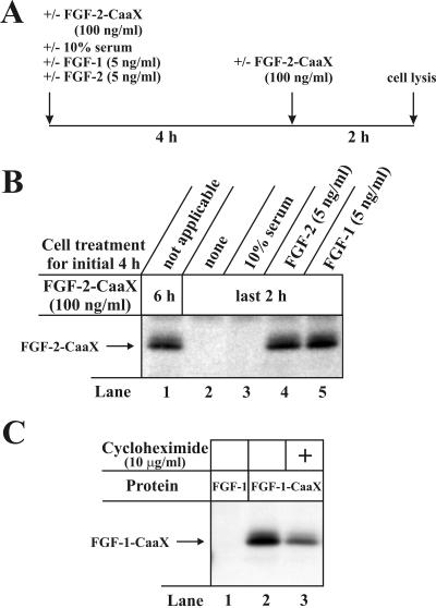
Figure 9.
FGF-2 translocation is induced by FGF-1 and FGF-2 treatment of NIH/3T3 cells. (A) Schematic representation of the time-course of the experiment. (B) Serum starved cells were preincubated with [14C]mevalonolactone, 1 μg/ml lovastatin and heparin. In one case 100 ng/ml FGF-2-CaaX was added for the entire 6 h incubation (lane 1). In other cases, the cells were for the initial 4 h left untreated (lane 2) or were treated with 10% serum (lane 3), 5 ng/ml FGF-2 (lane 4) or 5 ng/ml FGF-1 (lane 5) and then 100 ng/ml FGF-2-CaaX was added for the final 2 h of incubation. After lysis, cellular material was analyzed as in Figure 2A. (C) NIH/3T3 cells were pretreated as in Figure 2A and then treated for 6 h with heparin and either FGF-1 or FGF-1-CaaX in the absence (lanes 1 and 2) or presence of cycloheximide (lane 3). After lysis, cellular material was analyzed as in Figure 2A.

