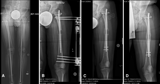
Fig. 1A–D.
(A) A preoperative radiograph obtained before LON shows the lower extremities of a 17-year-old male patient with a history of proximal focal femoral deficiency and a 3-cm leg length discrepancy on the left side. (B) A postoperative radiograph taken 4 weeks after surgery shows the distraction performed by the external rail. (C) At 8 weeks, the rail has been removed and the intramedullary nail has been locked distally to protect the well-formed regenerate bone. (D) At 5 months, the regenerate bone has fully consolidated. LON = lengthening over nail.

