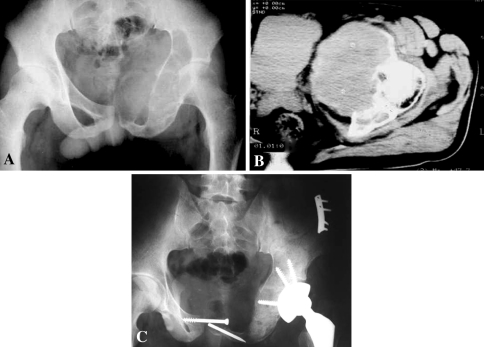
Fig. 1A–C.
(A) A plain film shows an osteolytic lesion in the left pubis, ischium, and periacetabular region of a 28-year-old female patient. This large lesion clearly involved the hip. (B) A CT scan shows the tumor involvement in the acetabulum where it produced a huge soft tissue mass. (C) The pelvic defect was reconstructed using recycled tumor bone implantation and a cemented THA was performed after tumor resection.

