
Figure 2. Transmission Electron Microscopy (TEM) of DEP and M.tb Uptake in Human Peripheral Blood Monocytes.
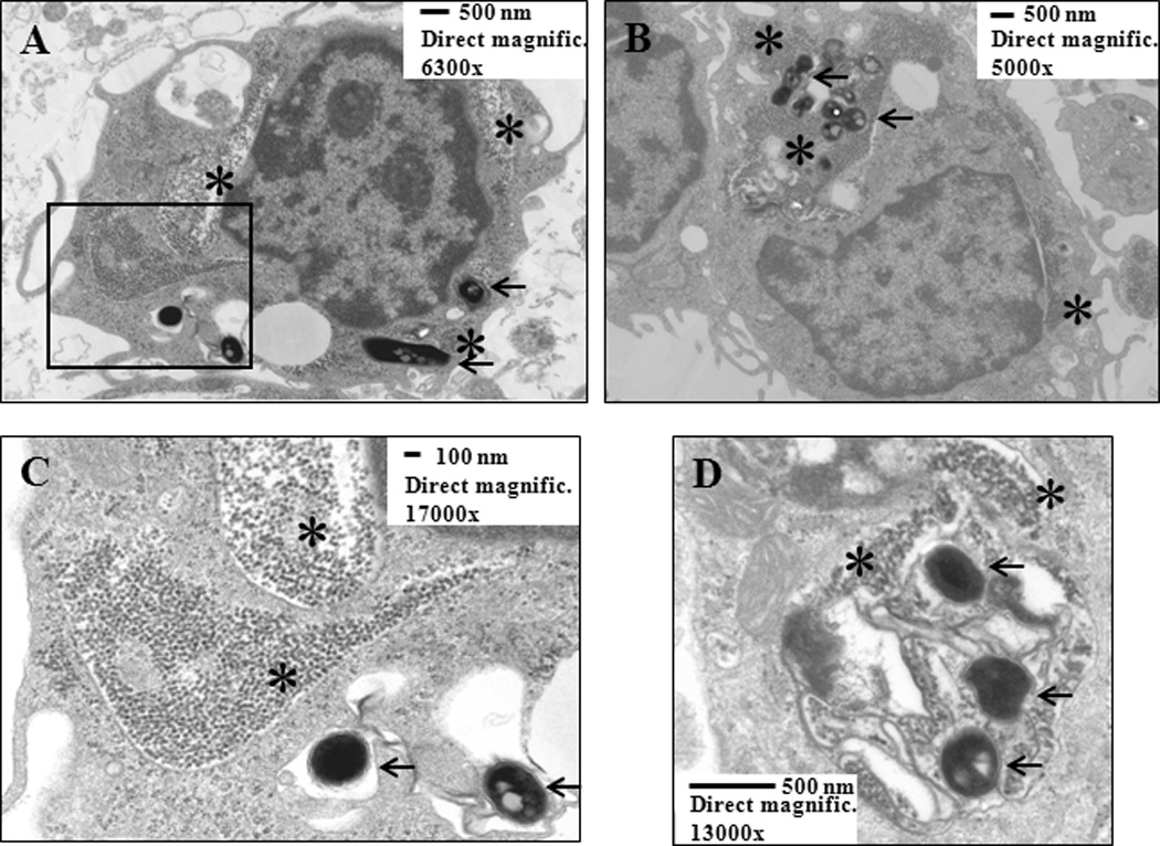
The micrographs (Panels 2A and B: overviews of two independent cells; 2C: details of the area surrounded by rectangle in 2A; 2D: intracellular compartment containing both DEP and three M.tb) show uptake of DEP (stars) and M.tb (arrows) within the same transects of monocytes, providing evidence of presence of DEP and M.tb within the same cell. Enriched peripheral blood CD14+CD3− monocytes were cultured with DEP (10 µg/mL) and M.tb (MOI 10) for 24 hours and processed as described in Materials and Methods. Scale bars and direct magnifications are included in the figure.