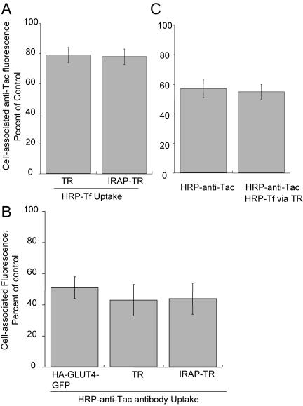
Figure 5.
Overlap of the insulin-regulated pathway with the furin pathway is restricted to TR-containing endosomes. (A) IRAP-TR, like TR, has access to only 20% of the Alexa488-anti-Tac fluorescence internalized by Tac-furin. Cells coexpressing Tacfurin and the TR or the IRAP-TR chimera were incubated with an Alexa488-labeled anti-Tac-antibody and HRP-Tf for 5 h. The cells were chilled to 4°C and treated with DAB/H2O2. The Alexa488 fluorescence in cells incubated with HRP-Tf is plotted as a percentage of the Alexa488 fluorescence in cells that were not incubated with HRP-Tf. The data are from a representative experiment and are the averages ± SEM of 25 cells per condition. (B) Fifty percent of the TR, the IRAP-TR chimera, and HA-GLUT4-GFP are in intracellular compartments accessible to Tac-furin. Cells expressing Tac-furin and the TR, IRAP-TR chimera, or HA-GLUT4-GFP were incubated with an HRP-anti-Tac antibody and Cy3-Tf (in cells expressing TR or IRAP-TR) or Cy3-anti-HA-antibody (in cells expressing HA-GLUT4-GFP) for 6 h. The Cy3 fluorescence is plotted as a percentage of the Cy3 fluorescence in cells that were not incubated with HRP-anti-Tac antibody. The data are the averages ± SEM of four independent experiments. (C) Tacfurin and HA-GLUT4-GFP overlap in TR endosomes. Cells expressing Tac-furin, TR and HA-GLUT4-GFP were incubated for 6.5 h in medium containing saturating concentrations of Cy3-anti-HA and HRP-anti-Tac antibodies with or without HRP-Tf. The cells were chilled to 4°C and treated with DAB/H2O2. The data are plotted as the percentage of Cy3 fluorescence in cells that were not incubated with either HRP-anti-Tac or HRP-Tf. The Cy3 fluorescence was divided by GFP per cell to correct for expression of HA-GLUT4-GFP. The data shown are the averages ± SEM (at least 20 cells per condition) from a representative experiment.

