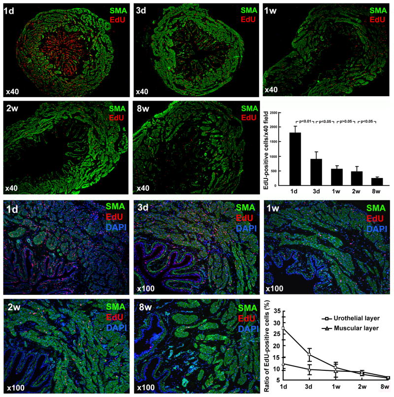
Figure 1.
EdU labeling and retaining in the developing bladder. Newborn rats were subject to intraperitoneal injection of EdU. Their urinary bladders were harvested at 1 day, 3 days, 1 week, 2 weeks, and 8 weeks later and processed for staining with SMA, EdU, and DAPI. Representative histological images at 40x and 100x are shown in the top and bottom two rows, respectively. The numbers of EdU-positive cells were counted at 40x magnification and presented in the bar chart. The ratios of EdU-positive cells (red stains) versus all cells (blue DAPI stain) were determined from the 100x photographs and presented in the line chart.

