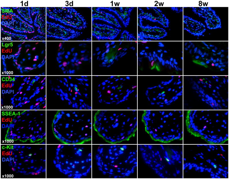
Figure 2.
Stem cell marker expression and EdU colocalization in the mucosa. Newborn rats were subject to intraperitoneal injection of EdU. Their urinary bladders were harvested at 1 day, 3 days, 1 week, 2 weeks, and 8 weeks later and processed for staining with SMA, EdU, and DAPI as well as the indicated stem cell marker (Lgr5, CD34, SSEA-1, or c-kit). Representative histological images of the mucosa at 400x and 1000x are shown for SMA (to row) and stem cell markers (bottom 4 rows), respectively.

