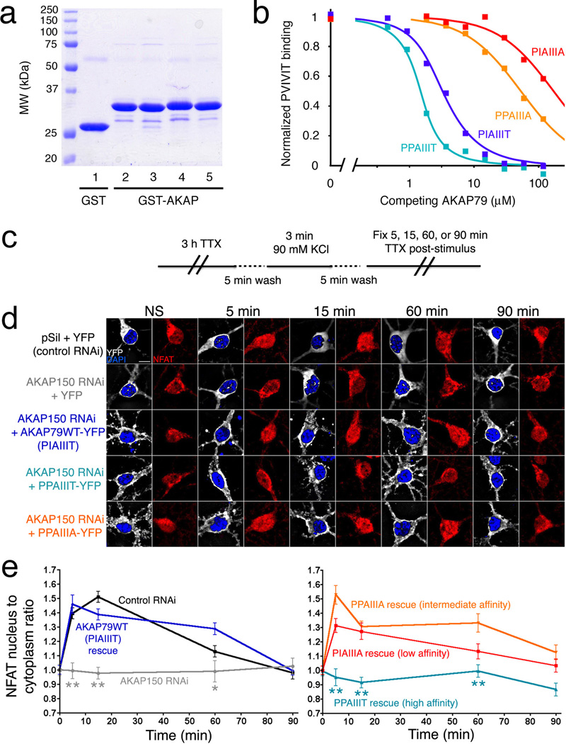
Figure 4.
A high-affinity AKAP79 variant, PPAIIIT, does not support NFAT nuclear translocation in hippocampal neurons. (a) Recombinant GST, GST-tagged wildtype AKAP79(333–408) (PIAIIIT, lane 2), and the variants PIAIIIA, PPAIIIA, and PPAIIIT (lanes 3–5) were analyzed by SDS-polyacrylamide gel electrophoresis and staining with Coomassie Brilliant Blue. (b) Kis estimated in a competitive binding assay are PPAIIIT, 0.08 µM; wildtype, 0.36 µM; PPAIIIA, 12 µM; and PIAIIIA, 39 µM. Total competitor concentration (not free concentration) is plotted. Data shown are representative of three experiments. (c) KCl stimulus protocol previously shown to activate L-type Ca2+ channel signaling through CN in hippocampal neurons21,29. (d) Summed intensity projection images of neuronal cell bodies and proximal dendrites in nonstimulated (NS) cultures and in cultures fixed at the indicated times after KCl stimulation. Transfection with control RNAi plasmid (pSil), AKAP150 RNAi plasmid, and RNAi-resistant expression plasmids is indicated. The paired images show YFP or AKAP-YFP (white), DAPI-stained nuclei (blue), and endogenous NFAT (red). (e) Time course of NFAT nuclear import after KCl stimulation, from experiments as in panel d, quantified as nucleus-to-cytoplasm mean fluorescence intensity ratios21. Each point represents n=12–25 neurons, and in each case the data have been normalized to the value for nonstimulated cultures (t = 0 min). Statistical comparisons were by one-way ANOVA with a Bonferroni post-hoc test, *p<0.05 and **p<0.01 compared to AKAP79WT rescue.

