Figure 6.
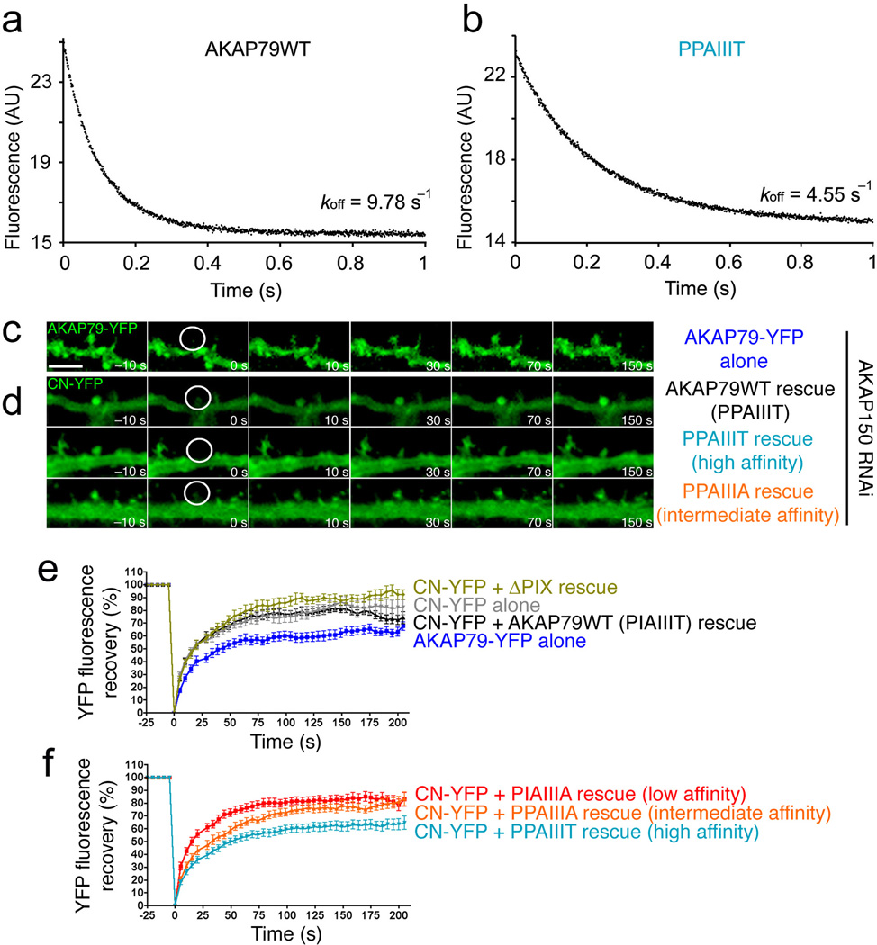
The high-affinity AKAP79 PPAIIIT variant decreases the rate of CN dissociation in vitro and reduces the mobility of CN in dendritic spines. (a) and (b) Dissociation of CN from (a) wildtype AKAP79(333–408) and (b) its I338P variant at 36 °C. (c) Time-lapse images of wildtype AKAP79-YFP fluorescence recovery in single dendritic spines of rat hippocampal neurons in culture at the indicated times after photobleaching. A prebleach image (t = −10 s) is shown for comparison. (d) YFP fluorescence recovery in neurons cotransfected with CN A-YFP (CN-YFP) and CFP-tagged wildtype AKAP79 (AKAP79WT) or CFP-tagged AKAP79 variants (PPAIIIT, PPAIIIA) as indicated. CFP fluorescence is not shown. (e) and (f) Percent YFP fluorescence recovery plotted against time for the FRAP experiments illustrated in panels c and d and for similar experiments with the other AKAP79 variants PIAIIIA and ΔPIX (deletion of PIAIIIT). In the experiment with CN-YFP alone, transfection with the RNAi plasmid was omitted. Maximal percent recovery (mobile fraction) values stated in the text were calculated from the curves in panels e and f by fitting a single exponential function. Statistical p values stated in the text were determined by one-way ANOVA with a Bonferonni post-hoc test (n=10–28 neurons).

