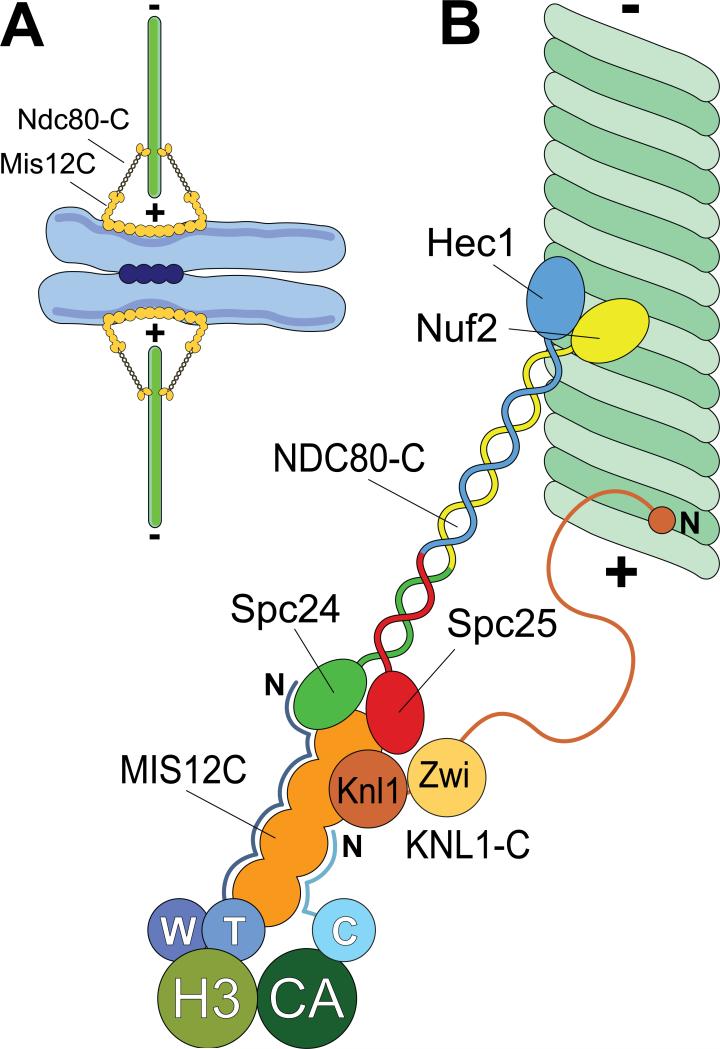
Figure 1. Schematic view of kinetochores.
A) During mitosis, sister chromatids are held together at centromeres by a cohesion complex (dark blue circles). Kinetochores (orange) assemble on centrometic chromatin and create a contact with microtubules (green). The plus (+) and minus (-) ends of microtubules are indicated. B) A close-up of kinetochores showing some of its components required for end-on microtubule binding. With the exception of the CENP-T/W complex (abbreviated as W and T) and CENP-C (abbreviated as C), all subunits of the constitutive centromere associated network (CCAN) have been omitted. CENP-T/W associates with histone H3-containing nucleosomes (H3), whereas CENP-C associates with nucleosomes containing the H3 variant CENP-A. The N-terminal region of CENP-T is an extended, largely disordered polypeptide chain that makes contacts with the 4-subunit Mis12 complex (MIS12-C) and with NDC80-C [11]. The N-terminal region of CENP-C is probably also disordered and makes contacts with MIS12-C [9,10]. The Knl1 complex (KNL1-C), which comprises Knl1 and Zwint-1 (Zwi), might contain a microtubule-binding site in the N-terminal region of Knl1 [28,56]. The C-terminal region of Knl1 interacts directly with the MIS12-C [70]. The NDC80-C is a tetramer. The Spc24 and Spc25 subunits interact with the MIS12-C, whereas the Hec1/Ndc80 and Nuf2 subunits face the microtubule.

