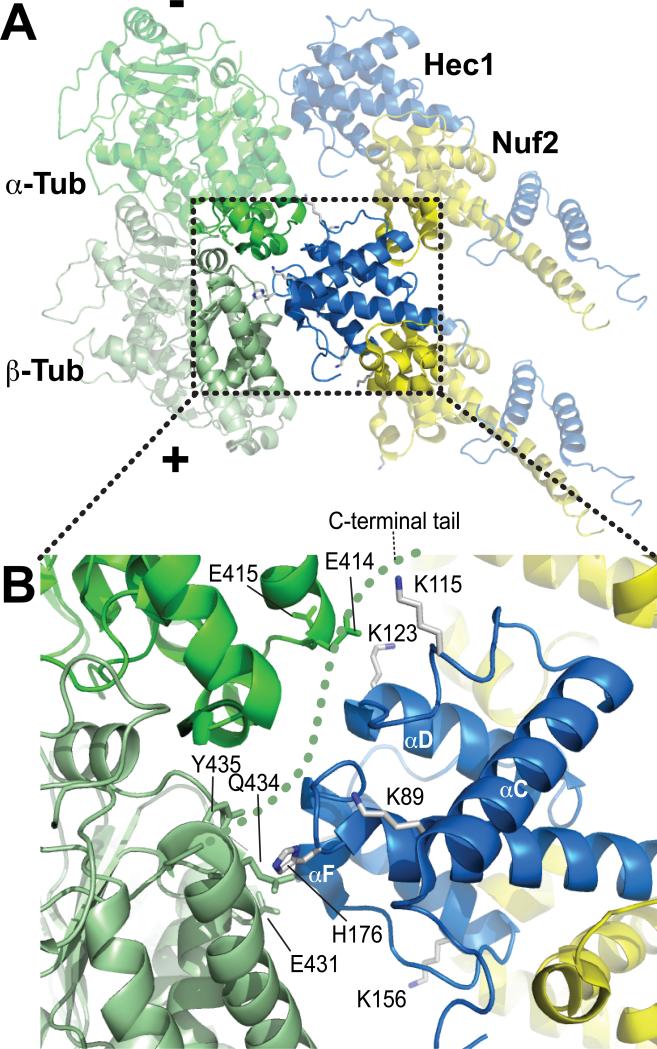
Figure 3. The “toe” and the “toe-print”, part II.
A) The view was rotated ~180° relative to the view in Fig. 2A. B) Close-up of the area boxed in A. The C-terminal tail of β-tubulin was invisible in the cryo-EM reconstructions. A hypothetical path for the C-terminal tail of tubulin (so-called E-hook) is shown as a dotted green line. K89 and K115 of Hec1 are potentially positioned for an interaction with the E-hook.

