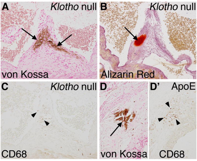
Figure 8. Macrophage infiltration is minimal in calcified lesions of Klotho null aortic valve leaflets.

Nodular calcification of the aortic valve hinge region (arrows) is apparent in a Klotho null aortic valve at 8 weeks of age as detected by von Kossa (A) and Alizarin red (B) staining. Macrophage infiltration was detected by immunoreactivity with CD68 visualized by brown DAB staining in the Klotho null valve (C). Sparse and isolated macrophages in the calcified region of the Klotho null aortic valve are indicated by arrowheads. High fat fed ApoE−/− mice were analyzed for inflammation associated with vascular calcification as a positive control. In the ApoE−/− aorta near the aortic root, calcification is apparent by von Kossa staining (D, arrow), in association with extensive inflammation, as indicated by CD68 staining (D′, arrowheads).
