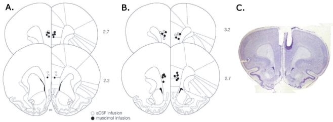
Fig. 2.
Reconstruction of microinjection sites. Cannula placements for muscimol and aCSF groups in Exp. 1 (A) and Exp. 2 (B) are depicted on illustrated coronal sections of mPFC (modified from Paxinos and Watson, 1998). Numbers on the right represent anterior-posterior distance from bregma. (C) Photograph of a representative coronal section showing the location of guide cannulae in the mPFC.

