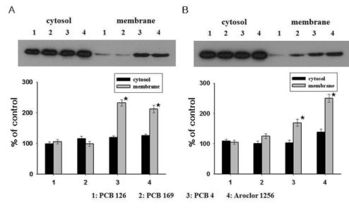
Fig. 2.
Translocation of cerebellar PKC isoforms from the cytosol to the membrane following PCBs exposure (50 µM) with western blot. (A) PKC-α made no apparent migration in PCB 126 or PCB 169 but made apparent migration from cytosol to membrane in PCB 4 and Aroclor 1256. (B) Since similar observation was made from PKC-βII, non-coplanar PCBs exerted greater effects on neuronal cells.
*Significantly different from control at p<0.05.

