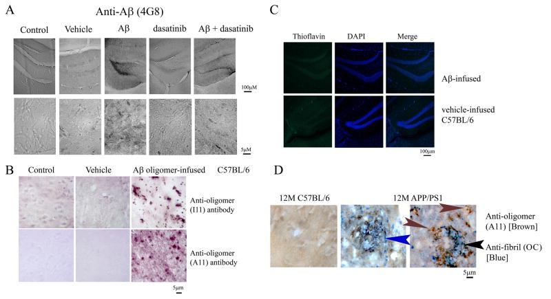
Figure 5. Brains of Aβ oligomer-infused animals were thioflavin negative but displayed immunoreactivity with anti-Aβ, 4G8, and anti-oligomer, A11 and I11 antibodies.
(A) Brains from wild-type control, vehicle-infused, and Aβ oligomer-infused animals (+/−) dasatinib were immunostained with anti-Aβ (4G8) antibody. Representative images from the dentate gyrus of the right hippocampus are shown. (B) Brains were also immunostained with anti-oligomer antibodies, A11 and I11, to detect infused peptide. Representative images of control, vehicle-infused, and Aβ oligomer-infused animals are shown. (C) Brains from vehicle-infused and Aβ oligomer- infused C57BL/6J mice were stained with thioflavin to identify the presence of any fibrils and counterstained with DAPI as a nuclear stain and Sudan black to quench autofluorescence. Representative images from the dentate gyrus are shown from 6 animals per group. (D) Brains from 12 month old C57BL/6J control and APP/PS1 mice were immunostained with A11 and OC antibodies to visualize fibrillar and oligomeric Aβ peptide deposition. Representative double label with A11 (brown) and OC (blue) is shown. Arrows indicate only A11 immunoreactivity (brown), only OC immunoreacitvity (blue) and co-localization (black).

