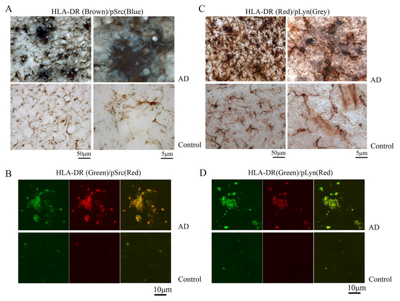
Figure 8. A population of AD brain microglia was immunoreactive for phospho-Lyn and phospho-Src.
AD and control temporal lobe sections were immunostained using anti-HLA-DR antibody to identify microglia, anti-phospho-Lyn-396 antibody to identify activated Lyn kinase and anti-phospho-Src antibody to identify activated Src kinase. (A) p-Src (blue)/HLA-DR (brown) double label and (C) p-Lyn (grey)/HLA-DR (red) double label images are shown. Images are representative of three cases each. AD and Control temporal lobe sections were also double-labeled using immunoflourescence using (B) anti-HLA-DR (green) and anti-pSrc (red) antibodies or (D) anti-HLA-DR (green) and anti-pLyn (red) antibodies.

