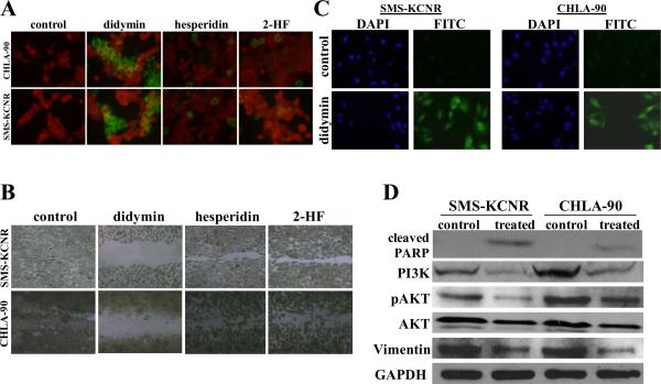
Figure 2. Pro-apoptotic and anti-migratory effects of didymin in neuroblastoma.
For TUNEL apoptosis assay, cells were grown on cover-slips and treated with 50 μM didymin for 24 h. TUNEL assay was performed using Promega fluorescence detection kit and examined using Zeiss LSM 510 META laser scanning fluorescence microscope with filters 520 and > 620 nm. Photographs taken at identical exposure at ×400 magnification are presented. Apoptotic cells showed green fluorescence (panel A). The in vitro migration assay shows that the didymin inhibits neuroblastoma cell migration in wound healing assay (panel B). For caspase activation detection, SMS-KCNR and CHLA-90 cells were plated on glass cover-slip in tissue culture treated 12-well plates and incubated with 50 μM of didymin for 24 h at 37 °C. Apoptotic cells were detected by staining with 5 μM Caspase FITC-VAD-FMK (Promega) in situ marker for 30 min (panel C). Analysis of the effect of didymin on PARP-cleavage, PI3K and Akt activation by Western-blot: The control and 50 μM didymin-treated cells were lysed and analyzed by Western-blot for PARP cleavage, PI3K (Y458/199), pAkt (S473), AKT and vimentin by using specific antibodies. Membranes were stripped and reprobed for GAPDH as a loading control (panel D).

