
Figure 1.
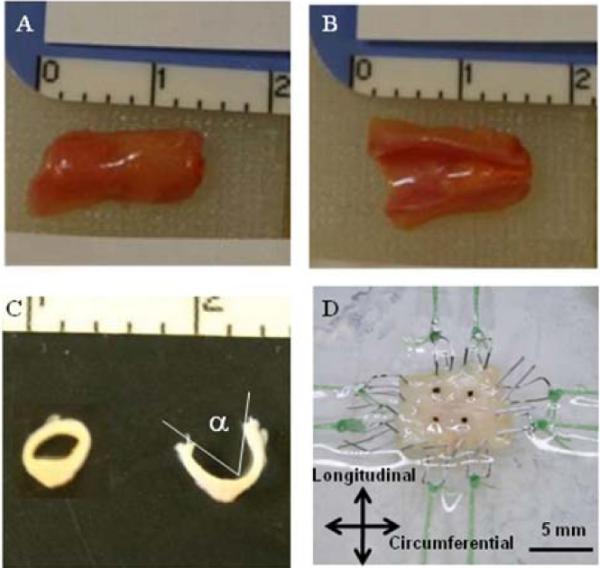
A coronary artery specimen before (A) and after (B) longitudinal cut, before (C inset) and after opening angle cut (C), and mounted on the biaxial testing machine via custom stainless steel hooks and tethers (D). Ruler scale is in centimeters in A, B, and C.