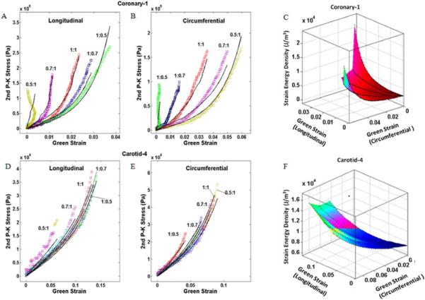
Figure 3.
Experimental data for a diseased coronary artery (Coronary-1) (A, B) and a carotid artery (Carotid-4) (D, E) with fit to Choi-Vito constitutive model. Strain energy density and the Green's strains along each axis for Coronary-1 (C) and Carotid-4 (F). Note the substantially larger spread in the curves typical for the coronary specimens indicating greater coupling between the axes than observed in the carotid specimens.

