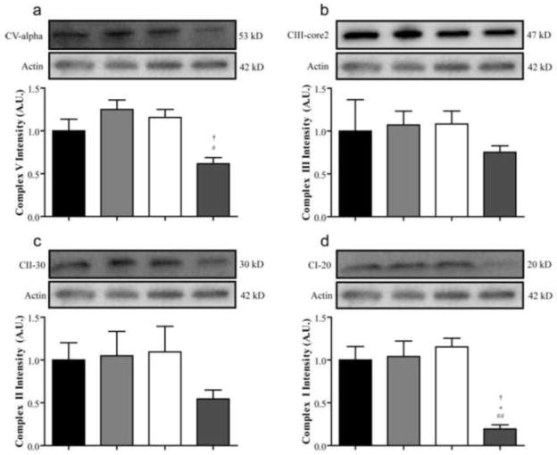
Figure 6. Mitochondrial oxidative phosphorylation proteins are reduced in diabetic BALB/cJ mice.
The expression of proteins from mitochondrial oxidative phosphorylation complexes was measured in the DRG after six weeks of diabetes. The expression of structural components of complexes V (a), III (b), II (c), and I (a) were all accessed by Western blot in nondiabetic BALB/cByJ (black bars, n = 3), diabetic BALB/cByJ (light gray bars, n = 3), nondiabetic BALB/cJ (white bars, n = 3), and diabetic BALB/cJ (dark gray bars, n = 3). Expression of both ATP synthase subunit α and Complex I subunit NDUFB8 were significantly reduced in diabetic BALB/cJ mice unlike diabetic BALB/cByJ mice. #p < 0.05 vs. nondiabetic BALB/cJ. ##p < 0.01 vs. nondiabetic BALB/cJ. *p < 0.05 vs. nondiabetic BALB/cByJ. †p < 0.05 vs. diabetic BALB/cJ.

