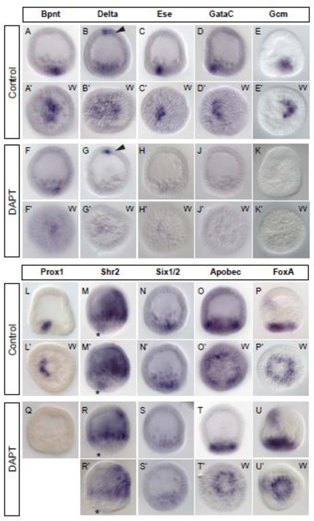
Figure 6.
Spatial effects of Delta/Notch perturbation with DAPT at 24 hpf. (A–N, Q–S) WMISH confirms quantitative results: Following DAPT treatment, mesodermal genes show either severely reduced or no staining by WMISH (bpnt, A/F; ese, D/H; gataC, D/J; gcm, E/K; prox1, L/Q; six1/2, N/S) . delta and shr2 are expressed in additional territories, but transcripts are specifically lost in the mesodermal domain (B/G: arrowheads indicate apical expression of delta; M/R: asterisk marks mesodermal expression of shr2). (O, P, T, U) The endodermal genes apobec and foxA do not clear from the cells that would normally be specified as mesoderm (compare vegetal view in O’/T’ and P’/U’), but the signal intensity remains the same. In lateral views apical is at the top. VV – vegetal view.

