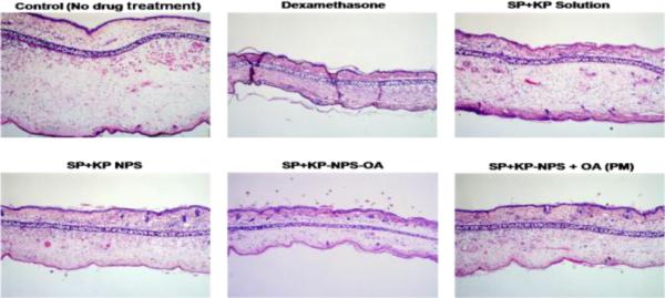
Figure 12.

H&E histological staining of ACD induced C57/BL mice ears after treatment with a positive control dexamethasone and a combination of two drugs SP and KP. The stained sections of mice ears after 72 h of treatment with SP+KP solution, SP+KP-NPS, SP+KPNPS-OA and SP+KP-NPS + OA (PM) are summarized in the figure. The images were taken using 10× lens.
