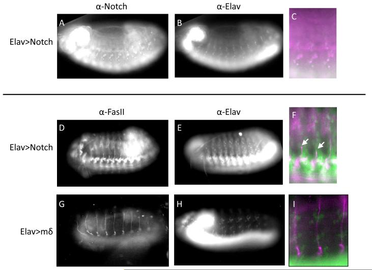
Figure 11. Notch pathway activation in neurons disrupts nerve outgrowth in embryos that is not induced with E(spl)mδ overexpression.
A-C) Embryos driving expression of the Notch intracellular domain (NICD), the active component of the Notch receptor, under control of elav show notch immunoreactivity in neurons. D-F) elav> NICD embryos stained for FasII (D) and elav (E) demonstrate stunted outgrowth of the ISN (arrow head). G-I) Embryos with targeted expression of the E(spl)mδ gene in neurons stained for FasII (G) and elav (H) show normal ISN outgrowth morphology.

