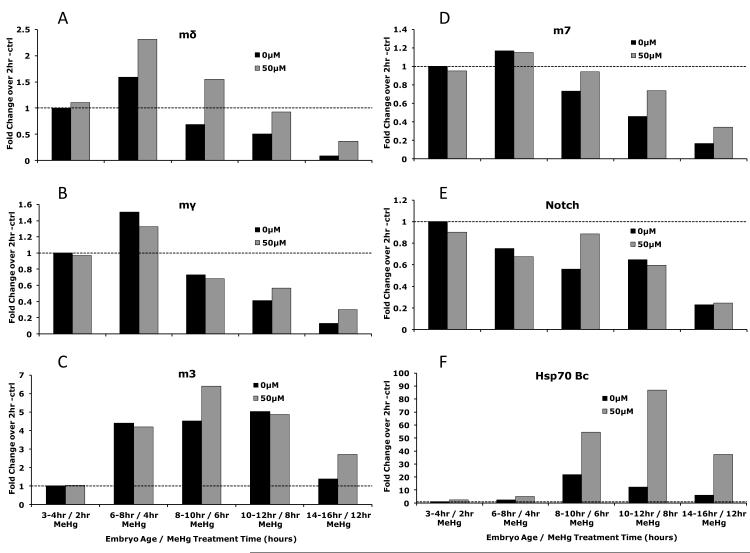
Figure 8. Response of E(spl) genes in Drosophila embryos after MeHg treatment over the course of development.
Drosophila embryos were dechorionated and soaked in buffer containing 50μM MeHg for indicated lengths of time. Gene expression determined by qRT-PCR is expressed in fold change over control treatments for A) E(spl)mδ, B) E(spl)mγ, C) E(spl)m3, D) E(spl)m7, E) Notch, and F) HSP70 Bc. (Each data point is derived from >300 pooled embryos from a treatment sampled at the indicated developmental time points).

