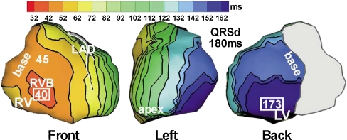
Fig. 3.
Epicardial isochrone maps during native rhythm in a patient with LBBB. Left anterior escending (LAD) coronary artery is shown and the approximate valve region is covered by gray. Earliest and latest ventricular activation times (in milliseconds) are indicated by framed numbers. Activation times are given with respect to QRS onset. QRSd QRS duration. Reproduced with permission [10]

