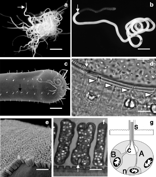
Fig. 1.

The stilbonematid nematode L. oneistus. a Light microscope image of an aggregation of approximately 50 living individuals; white arrow points to the beginning of the bacterial coat in one L. oneistus individual; scale bar is 2 mm. b Light microscope image of a single nematode; white arrow points to the beginning of the bacterial coat; scale bar is 150 μm. c Scanning electron microscope image of the anterior cuticle from which numerous setae protrude, black arrow points to one of them; scale bar is 20 μm. d Light microscope image of glandular sense organs (GSOs); the arrow points to the beginning of the bacterial coat, the arrowheads point to the canals of four GSOs; scale bar is 10 μm. e Scanning electron microscope image of the bacterial coat; scale bar is 3 μm. f Transmission electron microscope image of bacterial ectosymbionts; arrow points to one bacterium undergoing binary fission; scale bar is 0.5 μm. g Schematic representation of a GSO according to Bauer-Nebelsick et al. 1995 depicting its basic components: the A and B gland cells, the neuronal cell (n), the canal (C) and the seta (s). a and b are by Ulrich Dirks, c by Mario Schimak, d by Joerg A. Ott, e and f by Nikolaus Leisch
