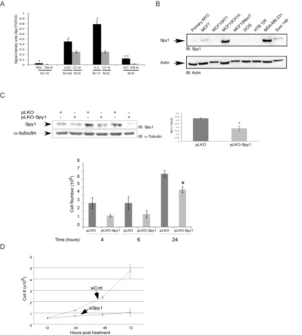
Figure 7.
Spy1 protein levels are elevated in human breast cancers.(A) TMAs containing cores from invasive ductal carcinoma (IvDC), infiltrated ductal carcinoma (IfDC), intraductal carcinoma (IDC) and invasive lobular carcinoma (ILC) as well as pair-matched normal (PM-N) or cancer-free patients (CF-N) were analyzed for Spy1 expression. The Spy1 signal intensity was normalized to nuclear stain (TOTO-3/PI) signal. Patient numbers are indicated below the sample (N). (B) Breast normal and cancer cell lines were analyzed by western blot analysis. Actin was used as a loading control. One representative blot of 2. (C) Western blot of 3 replicate infections of pLKO control or pLKO Spy1 in MDA-231 cells. Middle panel represents the densitometry values over 3 separate experiments. Where Spy1 is corrected for actin levels. Lower panel reflects cell counts at 4, 6, and 24 h post-infection. (All panels) Data shown is mean ± s.d. Student's t-test was performed * P < 0.05;**P < 0.01. (D) Knockdown effects on cell counts of MCF7 cells over 72 h after transfection with either pSUPER empty vector (siCntl) or pSUPER-Spy1 (siSpy1) over 3 separate transfections. Data shown is mean ± s.d.

