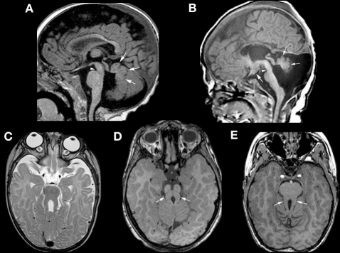
Figure 5.
Neuroimaging findings in molar tooth malformations. The characteristic imaging findings of a small vermis (small white arrows, A,B) and narrow isthmus (small white arrowhead, A,B) are identified on sagittal images. A tuber cinereum hamartoma is seen in (B). The variable appearances of the “molar tooth,” resulting from the large, horizontal superior cerebellar peduncles, are shown (white arrows in C, D, and E).

