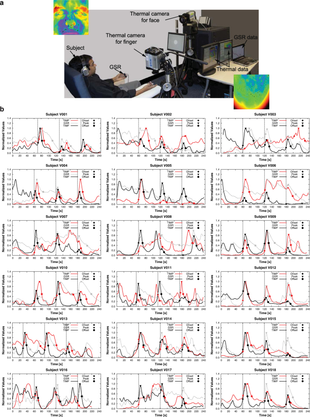
Figure 4.
(a) Lab experimental setup for validation of the perinasal sympathetic measurement via thermal imaging. The insets show snapshots of the subject's thermo-physiological responses on the perinasal and index finger areas following auditory startle. The black spots in the images indicate activated perspiration pores. (b) GSR, TIMF, and TIMP signals for all subjects in the validation data set.

