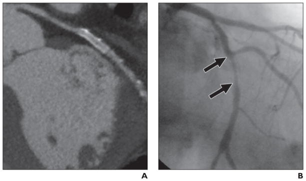
Fig. 4. 73-year-old symptomatic man with coronary artery disease who had drug-eluting 2.5 × 24 mm stent (Taxus, Boston Scientific) placed in left circumflex coronary artery 6 months and 10 days before studies. Image quality was good.
A, Qualitative and quantitative assessments by MDCT angiography did not show in-stent restenosis.
B, Quantitative coronary angiography shows no in-stent restenosis (arrows). Inset shows area of interest magnified.

