Figure 2.
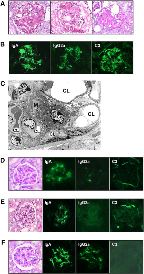
Histologic appearance and immune deposits in BALB/c mice implanted with 6-19 or 46-42 IgA RF–secreting cells and in immunoglobulin- or C3-deficient BALB/c mice implanted with 6-19 IgA RF–secreting cells. (A) Representative glomerular lesions of BALB/c mice implanted with 6-19 IgA RF transfectoma. Note segmental expansion of mesangial matrix and focal proliferation of mesangial cells (left), infiltrations of PMNs (center), and sclerotic changes (right) (periodic acid–Schiff staining; original magnification, ×400). (B) Immunohistochemical analysis of glomerular lesions of BALB/c mice implanted with 6-19 IgA RF transfectoma. Note fine granular deposits of IgA, IgG2a, and C3 in the mesangium (original magnification, ×400). (C) Electron microscopic findings of glomerular lesions in BALB/c mice implanted with 6-19 IgA RF transfectoma. Note prominent electron-dense deposits (indicated by white asterisks) within the mesangium (M). CL, capillary lumen; MC, mesangial cell (original magnification, ×1950). (D) Representative glomerular appearance and immune deposits in BALB/c mice implanted with 46-42 IgA RF hybridoma. Note essentially normal histologic appearance of glomeruli (periodic acid-Schiff staining) and only minimal extent of IgA, IgG2a, and C3 deposits in glomeruli (original magnification, ×400). (E) Representative glomerular appearance and immune deposits in immunoglobulin-deficient BALB/c mice implanted with 6-19 IgA RF transfectoma. Note essentially normal histologic appearance of glomeruli (periodic acid-Schiff staining) and the lack of C3 deposits (original magnification, ×400). (F) Representative glomerular appearance and immune deposits in C3-deficient BALB/c mice implanted with 6-19 IgA RF transfectoma. Note essentially normal histologic appearance of glomeruli (periodic acid-Schiff staining) despite substantial deposits of IgA and IgG2a (original magnification, ×400).

