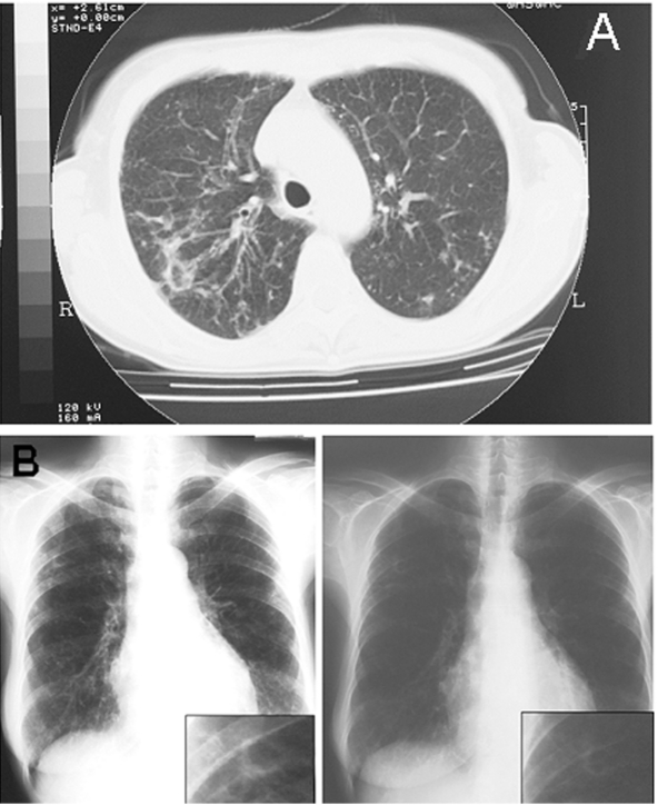
Figure.
A) Pulmonary computed tomographic scan representation of Mycobacterium lentiflavum lesions. Radiologic image shows the appearance of a widespread reticulonodular alteration and an opacity in the left middle lobe. B) Chest radiograph evolution after 3 months of treatment shows a sustained improvement of the radiologic alterations to the left pulmonary middle lobe.

