Fig. 7.
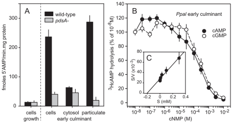
Localisation and substrate specificity of Ppal PdsA. (A) Stage specificity and localisation. Ppal wild-type or pdsA– cells were harvested at 0 hours (growth stage) or 16 hours of development (early culminants) and assayed for 3HcAMP hydrolysis as intact cells, or cells were lysed and assayed as separated soluble and particulate fractions. 5′AMP production was standardised on the protein content of the cell suspension or cell lysate. (B) Selectivity and affinity. Intact early culminant cells were incubated for 30 minutes with 10 nM 3HcAMP and increasing concentrations of cAMP or cGMP and assayed for 3HcAMP hydrolysis. Results are plotted as percentage of hydrolysis obtained at 10 nM 3HcAMP. (C) Hanes plot. Data from B were recalculated into absolute conversion rates (V) and plotted as a Hanes plot to estimate the Km of Ppal PdsA. All experiments represent mean and s.e.m. of at least two experiments performed in triplicate.

