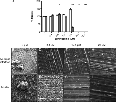
Fig 1.
Inhibition of biofilm formation on different surfaces by sphingosine. (A) L. monocytogenes biofilms were grown on polystyrene pegs and quantified using crystal violet staining. Planktonic growth at day 3 (□) and biofilm formation (▩), expressed as a percentage of control (n = 4, with the standard deviations [SD] shown). *, P < 0.01; **, P < 0.001; ***, P < 0.0001. (B to I) Representative SEM images of L. monocytogenes grown on food-grade stainless-steel coupons in the presence or absence of sphingosine at various concentrations. Images were captured near the air-liquid interface and the middle of coupons, where they were submerged in media. Bar, 10 μm. Magnification, ×5,000.

