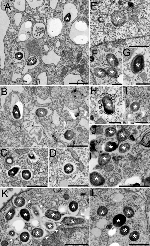
Fig 8.
Ultrastructural analysis of LVS and O-antigen-deficient LVS in human THP-1 cells in the absence of complement opsonization. (A and B) In the first 6 h after uptake of LVS bacteria, even in the absence of complement opsonization, the majority of LVS bacteria (indicated by asterisks) are found within membrane-bound vacuoles. (C to G) However, by 12 h after uptake (C and D), the majority of the LVS bacteria (asterisks) have escaped into the cytosol, while in the absence of serum opsonization, the majority of LVS ΔwbtDEF bacteria remain within membrane-bound vacuoles in the first 12 h after uptake (6 h [E] and12 h [F and G]). By 12 h after uptake, many of the LVS ΔwbtDEF phagosomes are decorated with a characteristic electron-dense fibrillar coat (faint in panel F and more obvious in panel G), which we also observed on the parental LVS phagosomes at earlier time points. (H) Blebbing and disruption of the fibrillar coat appear to accompany escape of the LVS ΔwbtDEF bacteria into the cytosol. (I and J) By 21 h after uptake, the majority of the LVS ΔwbtDEF bacteria have escaped and are free in the cytosol. (K and L) We observed similar features for the WbtIG191V mutant LVS. Panel L shows WbtIG191V mutant LVS bacteria at 16 h postinfection that have escaped into the cytosol. The wbtI-complemented strain (K) exhibits morphological interactions with the host cell similar to those of the LVS, whereas the WbtIG191V mutant strain (L) lacks the electron-lucent zone that is seen in the parental LVS. The electron-lucent zone surrounding the bacteria (K, arrowheads) is restored in the complemented strain. The experiment was conducted twice with similar results. Bars, 1 μm (A to E and J to L) and 0.5 μm (F to I).

