Fig 5.
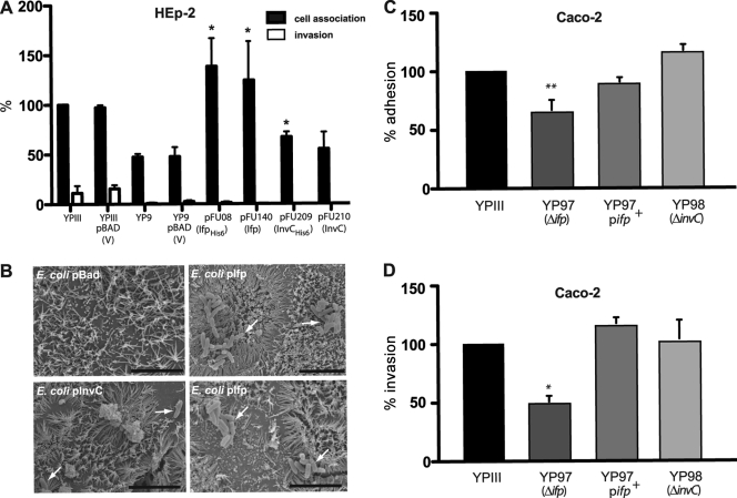
Ifp complements cell adhesion function of InvA. (A) Cell adhesion and invasion efficiencies of Y. pseudotuberculosis YP9 (ΔinvA) expressing Ifp (pFU08), IfpHis6 (pFU140), InvC (pFU209), or InvCHis6 (pFU210) were determined with human epithelial cells. Strains harboring the empty vector plasmid pBADmycA were used as a negative control. About 5 × 10⁶ bacteria were used to challenge 5 × 10⁴ cells, and the samples were incubated for 30 min at 20°C to determine cell attachment or at 37°C to monitor invasion efficiency. The data are presented as means ± standard deviations from three independent experiments performed in triplicate. Data were analyzed by the Student's t test. Asterisks indicate results that differed significantly from those for YPIII pBADmycA (P < 0.05). (B) E. coli K-12 cells harboring the empty vector pBAD-Myc, pFU140 (Ifp+), or pFU210 (InvC+) were used to infect polarized Caco-2 cells. Adherent bacteria were visualized by scanning electron microscopy and are indicated by white arrows. The black bars indicate 5 μm. (C and D) Monolayers of differentiated Caco-2 cells were challenged with YPIII, YP97 (Δifp), and YP98 (ΔinvC) and incubated for 3 h at 25 and 37°C to monitor cell attachment (C) and invasion (D). The total numbers of adherent and internalized bacteria were determined as described in Materials and Methods. The data represent the means ± standard deviations from three independent assays done in triplicate. Data were analyzed by the Student's t test. The asterisks indicate results that differed significantly from those for YPIII: *, P < 0.05; **, P < 0.01.

