Fig 3.
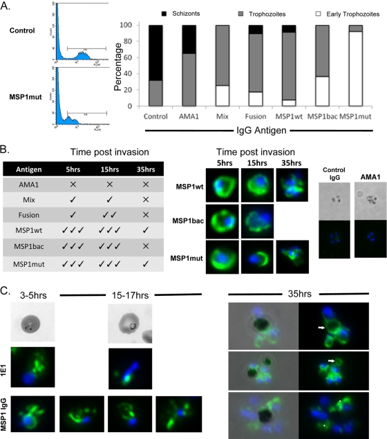
The effect of antibodies on intracellular parasite development. (A) Analysis of parasites ∼40 h postinvasion by FACS following hydroethidine staining and by microscopy following Giemsa staining. In the presence of specific antibodies, parasite development was delayed as shown by DNA fluorescence (ethidium labeling moved to left; MSP1mut antibody-treated parasites shown) and morphology compared to control parasites; the proportions of schizonts (black), trophozoites (gray), and early trophozoites (white) in each population are indicated in the graph. (B) MSP119-specific antibody is taken up into parasites at invasion and persists during development. The table on the left indicates the presence of detectable antibody at 5, 15, and 35 h postinvasion for parasites incubated with antibodies raised against the various proteins, and on the right are images of parasites from the different time points stained for the presence of rabbit IgG (green) and with DAPI (blue) to locate the nuclei. Representative images are shown for the groups that gave the most pronounced IgG labeling, along with the AMA1 and irrelevant IgG controls. (C) Comparison of MSP1 IgG labeling compared to control cultures containing MSP1-specific MAb 1E1 at early stages of parasite development, 3 to 5 and 15 to 17 h postinvasion. At later time points (35 h), MSP1 IgG labeling was evident and associated with hemozoin (arrows) and cytosolic structures (asterisks).

