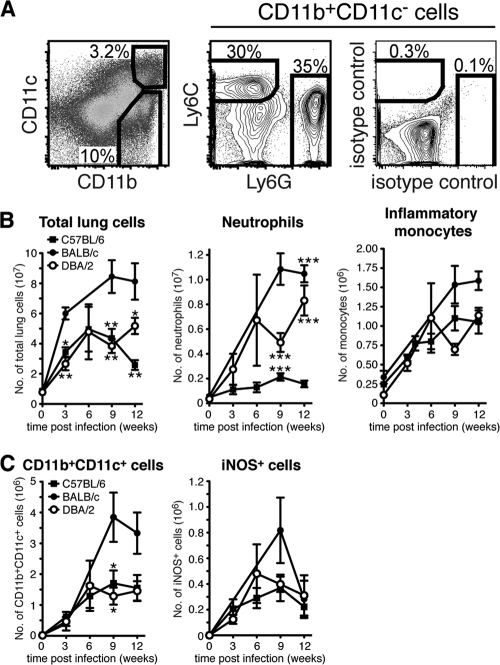
Fig 2.
Neutrophil, inflammatory monocyte, DC, and Mϕ populations in the lungs of naïve and M. tuberculosis-infected mice. (A) Single-cell suspensions were prepared from the uninfected or M. tuberculosis-infected lung tissues of individual mice and stained for cell surface expression of CD11b and CD11c (left), Ly6G and Ly6C (middle), or isotype control MAbs (right). Ly6C+ Ly6G− inflammatory monocytes and Ly6Cint Ly6G+ neutrophils were identified within the CD11b+ CD11c− gate (pregated on CD45.2+ CD19− cells [data not shown]). The plots show lung cells analyzed 12 weeks p.i. from one representative experiment. (B) Graphs displaying the absolute numbers of total lung cells (left), neutrophils (middle), and inflammatory monocytes (right) in uninfected lungs and at various time points after M. tuberculosis infection. (C) Graphs displaying the absolute numbers of CD11b+ CD11c+ cells (left) in uninfected lungs and at various time points after M. tuberculosis infection. iNOS-producing cells were identified in the CD11b+ CD11c+ subset (right). The absolute number of myeloid cells was determined in 3 to 11 separate experiments with 2 or 3 mice per group in each experiment. The results were pooled from all replicate experiments. The graphs show means ± standard errors of the mean (SEM). Statistically significant differences between the mouse strains are denoted as follows: *, P < 0.05; **, P < 0.01; ***, P < 0.001 by one-way ANOVA with Bonferroni posttest.

