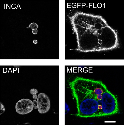
Fig 4.
EGFP-conjugated flotillin-1 localizes to C. pneumoniae inclusions. A549 cells were transfected with EGFP–flotillin-1 for 24 h and inoculated with C. pneumoniae for 48 h. Inclusions were detected with an antibody specific against IncA and anti-rabbit Alexa Fluor 647-conjugated secondary antibody. In the merged image, EGFP–flotillin-1 (EGFP-FLO1) is shown in green and IncA (INCA) in red. Bacterial and host DNA were stained with DAPI and are shown in blue. Bar, 10 μm.

