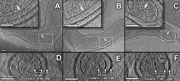
Fig 5.
Visualization of chemoreceptor arrays of Leptospira spp. Putative chemoreceptor arrays were observed in L. interrogans (A) and L. biflexa (B). The insets are the corresponding zoom-in views of the arrays outlined with white dashed lines. The arrays appear as clusters of pillar-like densities that extend from the IM and connect with a layer of high electron density at the membrane-distal ends. These are likely formed by CheA/W, which is known to form a continuous layer at the bottom of the chemoreceptor arrays. An additional array, unique to the saprophytic species, is found at the cell end (C). The inset is the zoom-in image of the array, indicating the presence of extra density layers (EL). The distance between the IM and the basal layer of CheA/W is relatively constant (25 nm) for most of the arrays, as shown in the cross sections (D and E). However, the novel array is longer (33 nm), as shown in the cross sections (F). The scale bar is 50 nm.

